
Advanced Diagnostic and Imaging Capabilities
Accurate diagnosis is the foundation of effective eye care. Modern imaging systems provide detailed views of ocular structures, allowing clinicians to identify abnormalities that may not be apparent during routine examination.
High-resolution imaging is particularly valuable for evaluating the retina, macula, optic nerve, and anterior segment. It also provides objective documentation that can be compared during follow-up visits.
Benefits
- Enables earlier detection of subtle pathological changes.
- Provides objective information for clinical decision-making.
- Supports accurate disease staging and monitoring.
- Helps clinicians evaluate treatment response over time.
- Creates valuable diagnostic records for long-term patient care.
Comprehensive Anterior Segment Evaluation
Detailed assessment of the cornea, anterior chamber, iris, and lens is essential for managing corneal disorders, cataracts, refractive problems, and contact-lens-related conditions.
Advanced anterior-segment assessment provides clinicians with a clearer understanding of ocular anatomy and helps identify structural abnormalities that can influence treatment decisions.
Benefits
- Improves assessment of corneal health and morphology.
- Supports evaluation of irregular corneal conditions.
- Helps determine suitability for different treatment approaches.
- Assists in contact lens and refractive management.
- Provides valuable information before surgical intervention.
Retinal and Macular Assessment
Retinal diseases can progress without noticeable symptoms during their early stages. Detailed retinal evaluation is therefore an essential component of a comprehensive eye hospital.
Advanced retinal assessment allows clinicians to evaluate retinal layers, macular structures, vascular changes, and optic nerve characteristics with a high degree of detail.
Benefits
- Supports early identification of retinal abnormalities.
- Helps monitor chronic retinal conditions.
- Enables precise comparison between follow-up examinations.
- Assists in determining appropriate treatment strategies.
- Helps protect vision through timely intervention.
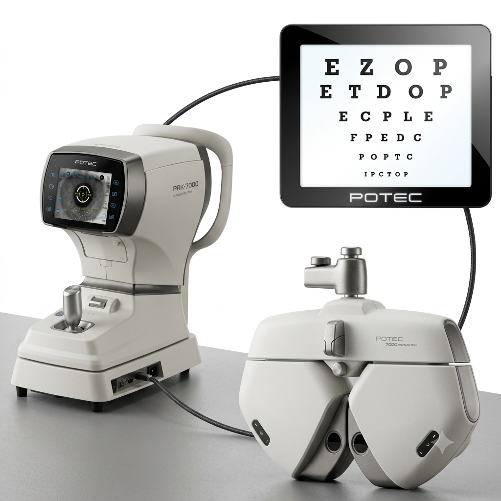
Precise Refractive Assessment
Accurate refractive evaluation is essential for providing appropriate vision correction. A comprehensive refractive assessment considers the patient's visual requirements while identifying changes in their optical status.
Precise measurements are particularly important when prescribing spectacles, contact lenses, specialty lenses, or planning refractive procedures.
Benefits
- Improves prescription accuracy.
- Supports personalized vision correction.
- Helps identify changes in refractive status.
- Improves patient comfort and visual performance.
- Provides reliable measurements for further clinical assessment.
Corneal Mapping and Structural Analysis
Detailed evaluation of corneal curvature and thickness is particularly important in specialty contact-lens practice, refractive assessment, corneal disease management, and surgical planning.
Understanding the three-dimensional characteristics of the cornea enables clinicians to make more informed decisions for patients with both regular and irregular corneal conditions.
Benefits
- Helps identify corneal irregularities.
- Supports specialty contact-lens fitting.
- Assists in evaluating patients for refractive procedures.
- Provides important information for monitoring corneal conditions.
- Improves the accuracy of individualized treatment planning.
Intraocular Pressure Assessment
Monitoring intraocular pressure is an important part of glaucoma evaluation and routine ophthalmic assessment. Reliable measurements can help identify patients who require further investigation and support long-term disease monitoring.
Benefits
- Supports early identification of abnormal pressure.
- Helps assess glaucoma risk.
- Enables monitoring of treatment effectiveness.
- Provides objective data for follow-up examinations.
- Supports long-term optic nerve protection strategies.
Functional Vision and Visual Field Assessment
Structural examination alone does not provide a complete picture of visual function. Assessment of the visual field helps determine whether a patient has functional deficits, particularly in conditions affecting the optic nerve and visual pathways.
Benefits
- Detects functional changes that may not be obvious to patients.
- Supports glaucoma assessment and monitoring.
- Helps evaluate progression of visual field loss.
- Provides objective information for treatment decisions.
- Enables comparison of functional changes over time.
Ocular Biometry and Surgical Measurements
Accurate ocular measurements are critical for procedures that require precise anatomical calculations. Reliable biometric assessment contributes to better surgical planning and helps clinicians select appropriate treatment parameters.
Benefits
- Improves precision during surgical planning.
- Supports appropriate intraocular lens selection.
- Reduces measurement-related uncertainty.
- Provides reproducible clinical data.
- Contributes to more predictable surgical outcomes.
Ophthalmic Surgical Infrastructure
A modern eye hospital should have a dedicated surgical environment designed around precision, sterility, patient safety, and efficient workflow. Surgical facilities should support both routine and specialized ophthalmic procedures according to the hospital's clinical services.
Benefits
- Provides a controlled environment for delicate procedures.
- Supports consistent surgical workflows.
- Improves patient safety and procedural efficiency.
- Helps surgical teams maintain high clinical standards.
- Enables hospitals to expand specialized ophthalmic services.
Conclusion
Building a modern eye hospital requires a strategic approach to equipment and infrastructure. The focus should not simply be on acquiring advanced technology, but on creating an integrated clinical environment where every capability contributes to better patient care.